Closing a wound looks like the final gesture of a procedure, but it is often the moment where a technically excellent surgery quietly loses ground. Sutures placed with the wrong material, tied under uneven tension, or spaced against tissue biology can turn clean approximation into inflammation, dehiscence, or lingering scar formation. Most of these problems are avoidable. The surgical literature has mapped the mechanics of tissue healing for decades, and the choices that lead to poor outcomes tend to repeat across clinicians and specialties in recognizable patterns. This article catalogs the most common suturing errors that undermine healing, explains why each one damages the tissue response, and outlines the concrete adjustments that turn closure into a reliable step rather than an unpredictable one. Whether the setting is oral surgery, dermatologic reconstruction, or general procedural work, the underlying principles remain constant, and the fixes are practical.
Choosing the Wrong Material for the Tissue
Suture selection is the first place errors accumulate. A material chosen out of habit rather than analysis creates avoidable friction with the healing process. Absorbable sutures in tissues that heal slowly are absorbed before the wound is structurally competent, and non-absorbable sutures in mucosal environments become chronic irritants that patients notice long after the procedure. Braided materials wick fluid and bacteria into the wound track more readily than monofilament equivalents, which changes the microbial exposure during the critical first days of healing.
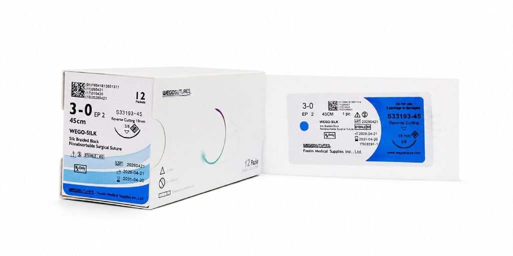
A useful mental model matches three variables at once: the tensile strength required over the healing window, the tissue’s inflammatory response to the specific material, and the practical handling qualities that let the operator place each stitch precisely. A well-chosen surgical suture respects all three constraints rather than optimizing for cost or convenience alone.
Tension Errors That Strangle Tissue
The most consequential suturing error is over-tightening. A knot pulled beyond gentle approximation compresses microcirculation at the wound edges, and the tissue responds with ischemia, edema, and sometimes frank necrosis along the suture line. The visible result is a raised, blanched, or discolored margin during the first postoperative week, followed by delayed healing and prominent scarring after resolution. Under-tightening produces its own problems, leaving wound edges apart long enough for granulation tissue to fill the gap and mature into a wide scar.
Learning the Correct Tension
Proper suture tension approximates the wound edges without indenting the tissue. When the knot is complete, the tissue on either side should sit lightly against its counterpart, with color and turgor unchanged. Reviewing wound tension immediately after each stitch — before moving on — catches the error while correction is still simple. A useful discipline is to place each stitch, tie loosely, evaluate approximation, and only then complete the throws that lock the knot.
Distributing Load Across the Closure
Concentrating tension on the middle stitch of a long incision guarantees that particular stitch will fail or cause local necrosis. Distributing load evenly across the entire suture line — with slightly closer spacing where mechanical stress is highest — spreads mechanical demand and preserves circulation. In tissues under skin tension, deep buried sutures that carry structural load allow the superficial closure to focus on precise edge alignment rather than mechanical support.
Poor Needle Handling and Track Trauma
The path of the needle through tissue creates a permanent record. A needle driven through tissue at an oblique angle enlarges the entry track and inverts wound edges, producing a subtly stepped closure that heals unevenly. Multiple passes through the same tissue — often the result of a first pass that missed the intended depth — create parallel tracks that macerate the wound margin and increase infection risk.
Refined technique treats each pass as a single deliberate motion: the needle enters perpendicular to the tissue surface, follows the curve of the needle body through both edges, and exits at a matched depth on the opposite side. When the geometry is wrong on the first pass, the correct response is to remove the needle, reassess, and re-enter cleanly rather than compensate through the same track.
Knot Errors That Loosen or Break
A well-placed suture undone by a poor knot is a fully preventable failure. Common knot errors include insufficient throws for the chosen material, twisting the knot rather than laying flat throws, and inadvertently backing off the previous throw while completing the next one. Different suture materials require different knot configurations to hold securely — monofilament materials generally need more throws than braided equivalents because of their lower coefficient of friction.
Practicing knot mechanics on training pads before applying them to patients produces measurable gains. Clinicians who work through knot drills quarterly maintain consistency across the volume of procedures they perform annually. Manufacturers such as Plovio publish knot-throw recommendations for each suture material in their catalog, and following those recommendations avoids the trial-and-error approach that leads to loose or breaking knots.
Ignoring the Timing of Removal
Non-absorbable sutures left in place too long leave stitch mark scars — the classic railroad-track appearance that patients associate with poor surgical outcomes. Removed too early, wound edges separate under mechanical stress. Location-specific timing matters: facial closures typically come out within five to seven days, back and extremity closures require longer intervals, and mucosal closures follow yet another schedule. Building removal timing into the postoperative plan at the moment of closure, rather than at the follow-up visit, prevents drift toward either extreme. When absorbable sutures are used, choosing a material with an absorption profile matched to the tissue’s healing rate accomplishes the same discipline automatically.
Closure Discipline as an Outcome Multiplier
Sutures are among the least glamorous elements of a surgical procedure and often the most decisive for how a patient remembers the result. Material selection matched to tissue biology, tension calibrated to preserve microcirculation, needle paths that respect wound geometry, knots that reflect the specific material, and removal timing tied to healing biology together determine whether a technically strong procedure produces a technically strong outcome. None of these disciplines require extraordinary skill or unusual equipment; they require attention and habit. Suppliers such as Plovio provide the material range and reference guidance that make disciplined closure practical across a variety of case types. Clinicians who invest in that discipline see quieter recoveries, cleaner scars, and fewer avoidable complications across their entire practice.